An upper superior and a lower inferior. Medial surface of lateral pterygoid plate and palatine bone.
Lateral Pterygoid Muscle Earth S Lab
Protrusion depression medial movement of mandible.
. Slightly reduced range of opening deviations of mandible with opening altered occlusion my teeth dont fit together right tinnitus are common complaints associated with TrPs in this muscle. The smaller superficial head has its origin mainly from the maxillary tuberosity with some fibers arising from the pyramidal process of the palatine bone. Click to see full answer.
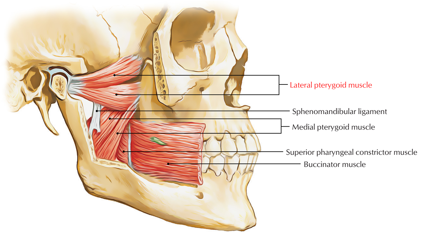
The medial pterygoid muscle attaches to the angle of the mandible and to the lateral pterygoid plate to form a sling with the masseter muscle that suspends the mandible Figure 6-19. The medial pterygoid is a muscle of your temporomandibular joint and can cause pain in the jaw throat and ear if it is tense or carries trigger points. Although having different origins both heads insert on the inner.
I usually save the lateral pterygoids for last when working on someones internal jaw muscles because they are harder to access. The medial pterygoid can be very hyperactive in jaw closing dystonia as a result of the so-called whack-a-mole phenomenon after repeated botulinum toxin injections into the masseter and temporalis muscles. The medial pterygoid muscle consists of two heads.
Your lateral pterygoid is a small thick muscle found on both sides of your skull just below the temples. Lateral pterygoid TrPs Refer often severe pain into the maxillary sinus deeply into the TM joint produce dysfunctions of the joint. It arises from the medial surface of the lateral pterygoid plate and the grooved surface of the pyramidal process of the palatine bone.
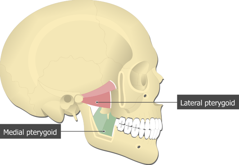
This muscle lies medial to the lateral pterygoid muscle. The lateral pterygoid muscle via its lower head separates the two distinct heads. It can assist in protrusion of the mandible.
The bulk of the muscle arises as a deep head from just above the medial surface of the lateral pterygoid plate. A Novel Treatment for Orofacial Pain Med Ultrason. Pterygoid branches of the maxillary artery.
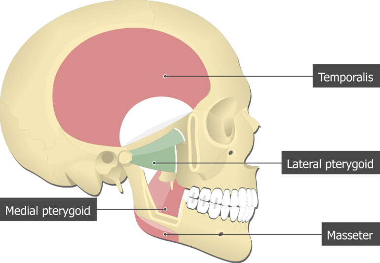
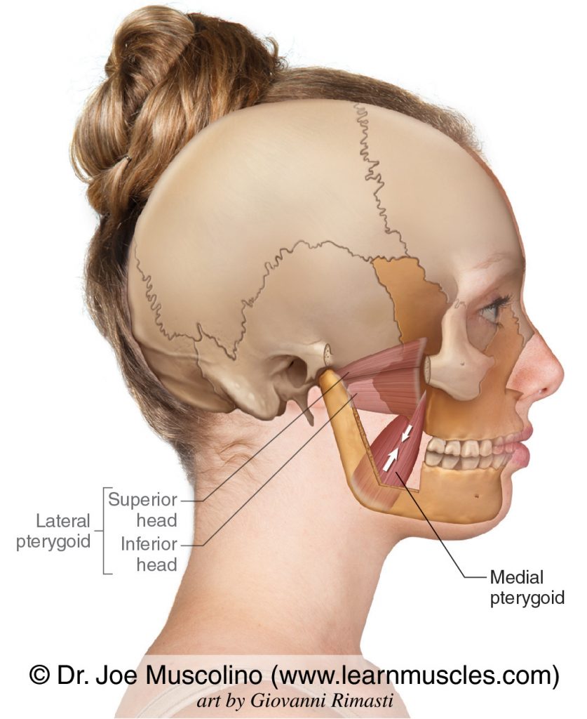
These two nerves pass between the medial and lateral pterygoid muscles. It belongs to the group of masticatory muscles along with the lateral pterygoid masseter and temporal muscles. The lateral pterygoid muscle is composed of two heads - superior and inferior.
These two nerves pass between the medial and lateral pterygoid muscles. The medial pterygoid muscle a major elevator of the jaw is a square-shaped masticatory muscle located on the medial aspect of the lower jaw bilaterally. The medial and lateral pterygoids - pronounced.
Both of the heads of your lateral pterygoid attach to the mandibular condyle. This muscle functions to move the lower jaw forward down and side. The superficial head is attached to the maxillary tuberosity and the pyramidal process of the palatine bone.
The primary action is to elevate the mandible and laterally deviate it to the opposite side. This muscle has two heads that originate from sections of the skulls sphenoid bone which has several other projected muscles. The lateral pterygoid is a short thick muscle somewhat conical in form which extends almost horizontally posteriorly and laterally between the infratemporal fossa and the condyle of the mandible.
It has two heads of origin. Ultrasound Guided Injection for Medial and Lateral Pterygoid Muscles. The smaller superficial head originates from the maxillary tuberosity and the pyramidal process of the palatine bone.
The nerve and artery usually pass between the two heads of the lateral pterygoid muscle. Easiest way to Memorize and remember both Pterygoid function. The medial pterygoid muscle is a thick and square shaped muscle.
The deep head of the muscle originates from the medial surface of the lateral pterygoid plate of the sphenoid bone. Jaw opening jaw deviation and jaw protrusion types of oromamdibular dystonia are caused by involuntary contraction of the lateral pterygoid muscles. The deep head is the major component and is attached to the medial aspect of the lateral pterygoid plate of the sphenoid bone.
Lateral pterygoid medial pterygoid. Thus the wide opening of the mandible may temporarily result in the mandible undergoing distortion in the transverse plane. The medial pterygoid muscle has a superficial and a deep head which arise from different areas of the jaw.
The lateral pterygoid Latin. It arises by two heads. Using the medial and lateral pterygoid muscles as references identify the buccal branch of the mandibular nerve CN V3 and accompanying buccal artery.
Branches of the mandibular division of the trigeminal nerve CN V3 Blood supply. It has a second slip of origin from the lateral surfaces of the pyramidal process of the palatine and tuberosity of the maxilla. Medial pterygoid muscle consists of two heads.
Ad Over 27000 video lessons and other resources youre guaranteed to find what you need. Medial pterygoid is a thick quadrilateral muscle that connects the mandible with maxilla sphenoid and palatine bones. The origin of both the bellies of the lateral pterygoid muscle is medial to their insertions.
The lateral pterygoid muscle is a small thick muscle located on each side of the skull that assists with mastication chewing. Musculus pterygoideus lateralis is a thick and short triangular-shaped muscle located in the infratemporal fossa of the skull. Ive found tension in the temporalises trigger points in the masseters and taut bands in the medial pterygoids.
It is also known as internal pterygoid muscle. Terra-goids These two assist the massester and temporalis muscles in movement of the jaw bone aka mandible. The medial pterygoid muscle has a triple function.
The medial pterygoid is a thick quadrilateral muscle. It helps to have tiny pinky fingers and even then sometimes I need to ask a client to shift their. On this page you will find instructions how to do this.
The nerve and artery usually pass between the two heads of the lateral pterygoid muscle. Medial surface of ramus and angle of mandible. It is classified as one of the primary muscles of mastication.
Protrusion elevation medial movement of mandible. Attachments of Medial Pterygoid Muscle. However its possible to eliminate these points and tensions with a self-massage.
Authors Yunn Jy Chen 1 Pei-Hsuan Chang 2 Ke-Vin Chang 3 Wei-Ting Wu 4 Levent Özçakar 5 Affiliations 1 Department of Dentistry School of. The superficial head of the medial pterygoid muscle originates from the maxillary tuberosity of the maxilla and pyramidal process of the palatine bone. Pain Trigger Points.
Using the medial and lateral pterygoid muscles as references identify the buccal branch of the mandibular nerve CN V3 and accompanying buccal artery. Medial Lateral Pterygoids I almost forgot about these two lil jaw muscles.
![]()
Medial And Lateral Pterygoid Muscle Anatomy And Function Kenhub

Lateral Pterygoid Muscle Wikipedia
Lateral Pterygoid Muscle Attachments Actions Innervation
Medial Pterygoid Learn Muscles
Medial Pterygoid Muscle Attachments Actions Innervation
![]()
Medial And Lateral Pterygoid Muscle Anatomy And Function Kenhub
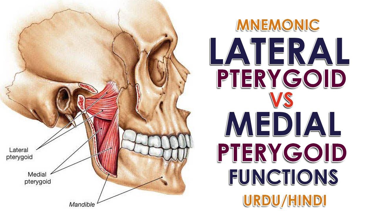
Mnemonic Lateral Pterygoid Vs Medial Pterygoid Function Urdu Hindi Youtube

Musclemonday Pterygoid Muscles Today We Are Going To Discuss Our Last Muscle Of The Week The Medial And Lateral Muscle Health And Wellness Physical Therapy
0 comments
Post a Comment